Resonance in Chromotherapy Treatments
Healing Leishmaniasis Infection by Red Light at 644 nm
by Alex Putney
January 26, 2018
As one of the most severe diseases known to humanity, Leishmania comprises a genus of flagellate protozoan parasites prevalent in tropical regions that are transmitted by the bite of infected female sand flies of the genera phlebotomus and Lutzomyia (Van der Meide et al., 2005).
Approximately 12 million people suffer from Leishmania infections as cutaneous, mucosal and/or visceral lesions. Mucosal Leishmaniasis is the most severe form of the infection, caused by the spread of parasites from cutaneous lesions to the mucosal membranes, where they progressively destroy connective tissues and cartilage throughout the nasopharyngeal tract (Tavares et al., 2003; Herwaldt, 1999).
Two medicines containing antimony are commonly prescribed for Leishmania infection: meglumine antimoniate (Glucantime) and sodium stibogluconate (Pentostam). However, these medications are painful and expensive, and increase the toxicity of the patient over the extended course of treaments. However, recent scientific findings have demonstrated the efficacy of chromotherapy against Leishmaniasis.
Effective photonic methods for healing of Leishmaniasis infections were first reported by Azeemi et al. in 2011. Despite the inexpensive and easy application of these new healing modalities, such findings have been largely ignored by media and subsequent scientific journal publications on the same subject. Quoted here at length, these outstanding results were reported as 'Effects of Different Colours in the Visible Region on Leishmania tropica' (Azeemi et al., 2011):
Abstract
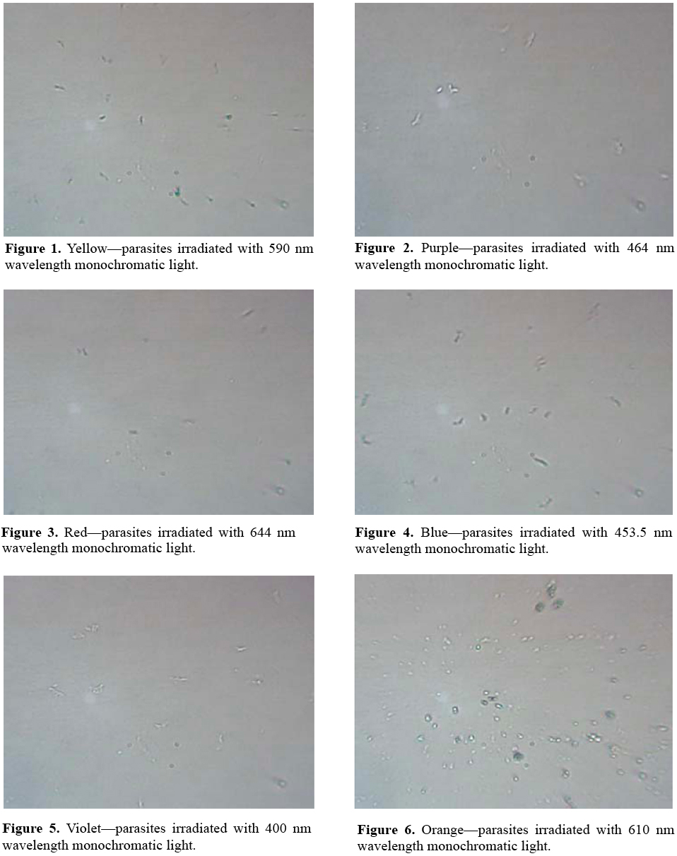
The aim of this study was to investigate the wavelength-dependency of chromotherapy effects on cutaneous Leishmaniasis parasite growth. Chromotherapy uses visible range radiations to improve healing; however, its effects on parasite are not well understood. Leishmania tropica was irradiated using seven (7) different wavelengths of visible region. Optical density was observed, which showed that red colour (644 nm) wavelength inhibited the growth of parasite while other colour wavelengths also affected the growth of parasite. It is, therefore, suggested that as red colour inhibits the growth of parasite so patients suffering from L. tropica can be treated with the application of red colour.
Results
After initial incubation [of Leishmania tropica] for production, out of six samples two were found positive and other four were contaminated with bacteria and fungus. Yellow (590 nm) and purple light (464 nm) increased the size of the parasite (Figures 1 and 2). Red (644 nm), blue (483.5 nm) and violet (400 nm) decreased growth considerably; the size as well as the number of counts of the parasite, with red the most effective (Figures 3-5). Orange colour (610 nm) increased growth incredibly while in green colour (538 nm) the promastigotes appeared to change their size and shape and somewhat converted into rounded form (Figures 5 and 6). In the sample irradiated with red colour wavelength, the parasites were observed as lethargic... as well as least number of counts was observed...
Discussion
It was discovered that Monochromatic Single-Wavelength Light Beams had an excellent therapeutic effect on afflicted cell tissue. This occurs through a process called "Photo-Stimulation." The low intensity (non-coagulative) visible laser radiation has been successfully used in some areas of medicine (photodynamic therapy of tumors, therapy of infant hyperbilirubinemia, some dermatological diseases, etc.) [Pratesi et al., 1980]. Also the therapy with red (632.8 nm) laser light (stimulation of tissue regeneration) used for irradiation of the patients with trophic and indolent wounds has gained acceptance in the clinical practice [Karu et al., 1984]. The biomodulatory effect can have a positive effect on the repair of cutaneous wounds [Mendez et al., 2004]. Various studies have been carried out that show the effect of monochromatic light on cells, but the research lacks empirical data regarding effects on parasite...
Figures 1-8 are self explanatory to reflect the image of optical density of Leishmaniasis. L. tropica when exposed to red light (644 nm), the decay in this case followed the Gaussian tail and finally to a ramp function. This is an indication that the cutaneous Leishmaniasis, after decay, just disappears because the parasites die...
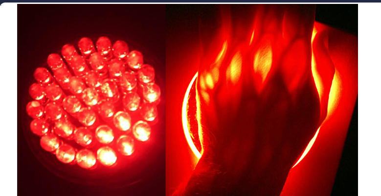
Although Low Level Laser Therapy (LLLT) has been used previously in most studies, coherence is not important when photo-biological effects are expected because both coherent and non-coherent light have been shown to be effective [Karu et al., 1987]. In our study it is observed that red light showed a great change to inhibit the growth of Leishmania tropica organisms. From this study it is evident that cutaneous Leishmaniasis can be eliminated with higher wavelength, i.e., at 644 nm and with least energy (filtered light beams).
Conclusion
Red color (644 nm) inhibits the growth and becomes responsible for the decay of leishmania parasite while orange color (610 nm) increases the growth of parasite. Undoubtedly this makes the procedure of chromotherapy for treatment of leishmaniasis cost effective and easy approachable. The response of Leshmania parasite to each color is unique and this confirms Chromotherapy (with 644 nm wavelength), to be very easily manageable by the patient with no problems during the treatment.
While these breakthrough findings conform to a rapidly expanding class of non-invasive photonic treatments that are cheaper, simpler and far more effective than conventional chemical-based medications, the exact physiological mechanisms behind their greater efficacy have not been clearly identified.
The authors of this straight-forward chromotherapy study characterized their findings as effects of 'photo-stimulation' that engages cellular regeneration processes in the body for enhanced wound healing. In the years following this study, investigations of the qi meridian system of the human body have successfully identified the production of new stem cells in the qi meridians that dramatically enhances cellular regeneration.
First conducted by Korean biophysicist Bonghan Kim and reported in 1965, Kim's findings have since been confirmed and expanded upon by dozens of other research teams. The structure of the qi meridian system is composed of Bonghan ducts (transparent channels), Bonghan corpuscles (duct junctures) with Bonghan granules, also called microcells or sanals, containing DNA fragments that trap ambient light as they flow through the channels.
The flow speed of DNA-containing granules within qi meridians on the surfaces of mammalian organs has been reported (Sung et al., 2008), while exposure to ultraviolet A (360 nm) light was shown to increase the flow rate of granules within the meridians in a report titled: 'UV-A Induced Activation of Bonghan Granules in Motion' (Sung et al., 2005). A subsequent paper identified the 'Bonghan System as Mesenchymal Stem Cell Niches and Pathways of Macrophages in Adipose Tissues' comprising the mechanism behind adipogenesis: the cellular differentiation process generating adipocytes (Lee et al., 2009).
A concise summary of diverse findings related to the light-enhanced regenerative effects of stem cell production by the qi meridian system was presented in a contemporary paper entitled 'Bonghan Circulatory System as an Extension of Acupuncture Meridians' (Soh, 2009):
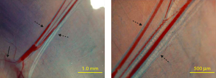
Trypan blue technique revealed Bonghan ducts (dotted arrows) along bundle of blood vessels and nerves; right panel, magnified view clearly showing duct along blood vessel; bundle of blood vessels and nerves connect tumor tissue (arrow) at lower left corner to outside skin (Yoo et al., 2009).
The Bonghan system is a newly-discovered circulatory system, which corresponds to classical acupuncture meridians and was discovered in the early 1960s by Bonghan Kim. Despite its potential importance in biology and medicine, it has been ignored or forgotten for a long time. Only recently have most of its significant parts, such as the Bonghan system inside blood or lymph vessels, on the surfaces of internal organs, and in brain ventricles, been confirmed...
A direct test to demonstrate liquid flow, performed by injecting fluorescent nanoparticles into an organ-surface Bonghan corpuscle, revealed a oneway flow, as expected for a circulation system (Lee et al., 2005). The average flow speed, recently measured by injecting Alcian blue into a Bonghan corpuscle on the surface of a rabbit liver (Sung et al., 2008), was 0.3 ± 0.1 mm/s, in agreement with Bonghan Kim's data (Kim, 1965). Liquid flow through a Bonghan duct from the skin toward the internal organs was observed by injecting chrome-hematoxylin and fluorescent nanoparticles in the skin near a rat testis...
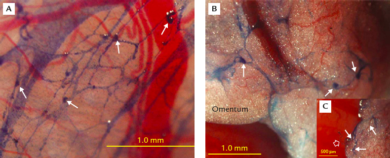
Weblike network of Bonghan ducts revealed by using trypan blue. (A) Web of Bonghan ducts on visceral peritoneum around stomach near rat sleen; several small Bonghan corpuscles at crossing points (arrows); blood capillaries not stained. (B) Network of Bongan ducts on omentum below stomach and over small intestine; threee small corpuscles at crossing points of Bonghan ducts (arrows). (C) Inset: another part of same omentum as (A); floating Bonghan duct (open arrow) connected to Bonghan ducts (arrows) in omentum, showing Bonghan ducts on omentum as part of larger network of freely movable Bonghan ducts on internal organ surfaces (Lee et al., 2007).
Improved immune function and beneficial effects on inflammation are often described after acupuncture treatment (Son et al., 2002) and an abundance of mast cells is reported at acupuncture points (Hwang, 1992). We observed that the organ-surface Bonghan corpuscle and Bonghan duct contained a significant number of monocytes, eosinophils, mast cells, and macrophages (Lee et al., 2007; Yoo et al., 2007; Ogay et al., 2009). The abundance of such immune cells in the Bonghan duct supported evidence for the related therapeutic effects of acupuncture treatment and for Bonghan Kim's claim that the organ-surface Bonghan duct is an extension of the classical acupuncture meridian system.
Blood cells are known to be generated in the bone marrow but Bonghan Kim claimed that the intravascular Bonghan duct is another hematopoietic organ (Kim, 1965). Indeed, we observed here that the Bonghan duct became thicker and thus easier to detect when anemia was induced by the injection of phenylhydrazine. Many red blood cells in early stages of maturation were observed in organ-surface Bonghan corpuscles when anemia was induced.
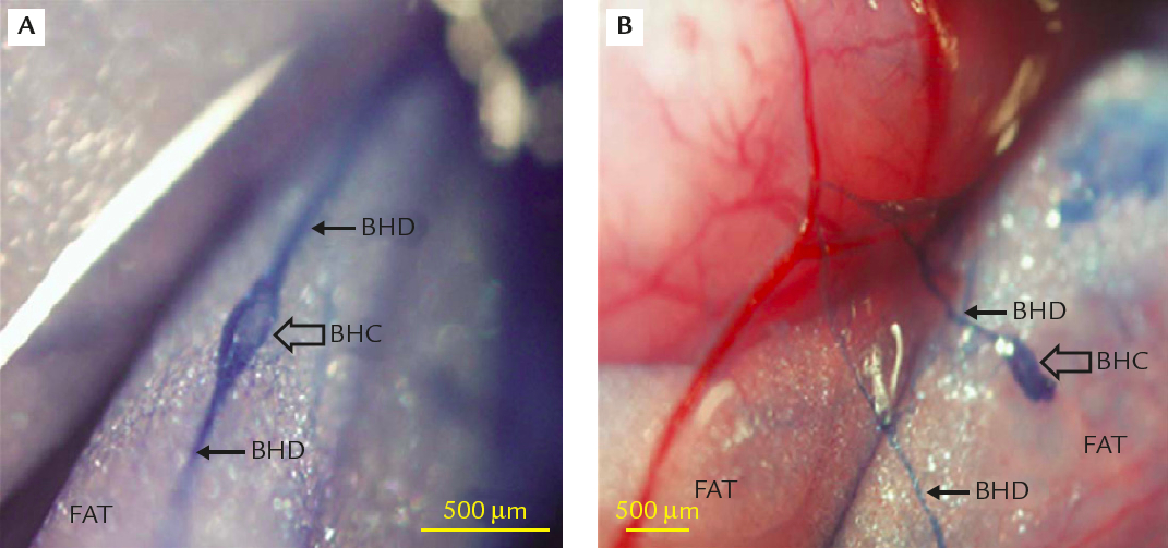
Trypan blue staining of Bonghan duct and Bonghan corpuscle inside adipose tissues. (A) Bonghan corpuscle and connected Bonghan duct inside adipose tissue around rat small intestine. (B) Bonghan corpuscle and two Bonghan ducts near same rat small intestine; blood vessels and adopise tissues not stained (Lee et al., 2009).
Regeneration of damaged liver cells was reported in Bonghan Kim's fourth article (Kim, 1965). Considering this claim, we hypothesized that there might be adult stem cells in Bonghan corpuscles and to verify this hypothesis, we stained sliced Bonghan corpuscles and Bonghan ducts with stem cell marker antibodies. We observed that mesenchymal stem cell markers were strongly expressed in a manner similar to bone marrow...
[Specific] protein profiles suggested that Bonghan Ducts located on organ surfaces have roles as temporary depots and points of differentiation of stem cells for tissue regeneration. Damaged liver tissues are regenerated by the gathering of sanals that had migrated through the Bonghan ducts (Kim, 1965). This process has not been specifically investigated here, but some basic studies have been performed on Bonghan microcells, revealing that their motion appeared to be Brownian, but that they also showed some peculiar light interactions. Their average speed was not affected by visible light, but was significantly increased by UV-A (360 nm) (Sung et al., 2005). The presence of DNA inside a sanal was identified using various types of DNA-specific staining,... and the state of the DNA shown to be fragmented... (Ogay et al., 2006).
These remarkable findings concerning the structural features and flow dynamics of the qi meridian system reveal the complete mechanism by which tissue regeneration is stimulated by exposure to red light (644 nm), and more significantly enhanced by exposure to ultaviolet A light (360 nm), whereby mesenchymal stem cells are produced, distributed and assembled at wound sites requiring regeneration.
This extremely well supported conclusion informs a paradigm-shifting reinterpretation of chromotherapy as an effective method for achieving stem cell regeneration treatments that cost thousands of times less than present-day stem cell injection methods, by directly stimulating the qi meridian system itself.
Irradiation of specific acupressure nodes along the qi meridians by traditional Chinese medicine moxa (infrared) treatment has been extensively studied through biophotonic imaging processes, revealing electro-stimulation of nodal points significantly enhances known acupressure and acupuncture modalities.
Through the use of ancient Atlantean healing techniques, still practiced in secret in various parts of the world to this day, Qigong and Mo Pai practitioners such as John Chang of Java, Indonesia are able to apply electrical currents to acupressure needles by consciously focusing qi energy stores in their own bodies, for emission through their bare hands. Chang can even ignite paper with his palms.
Synthesis of ancient healing modalities with modern medical breakthroughs offers not only the alleviation of disease factors in the body, but furthermore significantly enhances the human lifespan by cellular regeneration through the process of autophagy, whereby damaged organelles within cells are repaired and the aging process is reversed. Through these ancient means, newly rediscovered, the mystical pursuit of enhanced longevity finds its fulfillment.
Copyright 2018 Alexander R. Putney